Myelography and Myelogram
Myelography and Myelogram
Overview
A Myelography (also called a Myelogram) is an outpatient diagnostic procedure to examine the spine. It allows doctors to identify problems involving the spine, the spinal cord, and the nerve roots that are often not clearly revealed with x-rays, MRIs, or CT scans.
Preparation
To begin, you lie on your stomach. Your back is cleaned with an antiseptic. Local anesthesia is used to numb the skin and tissue of your back.
Injecting the Dye
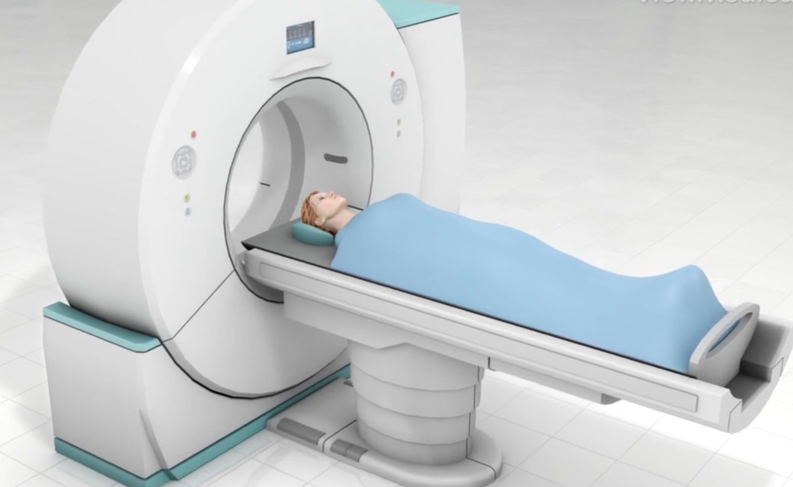
The physician carefully guides a different needle down to the spinal column and through the thin sac (dura) that surrounds your spinal cord and nerve roots. Once the needle is correctly positioned, the doctor slowly injects contrast dye to mix with your spinal fluid. The doctor will then use an x-ray or CT scanner to take images of your spine, with the dye making certain anatomy more clearly visible.
End of Procedure
When the exam is complete, your injection site is bandaged. You will be briefly monitored before being discharged to go home. You will not know the results of the exam when you leave. A radiologist will evaluate the study and give your doctor a report of the findings. You will learn the results from your doctor at a follow-up visit. In the meantime, your body will harmlessly eliminate the dye.
Revised from www.viewmedica.com © Swarm Interactive. Unauthorized duplication is strictly forbidden.
- Category / Diagnosis


