Anatomy of the Knee
Anatomy of the Knee
Overview
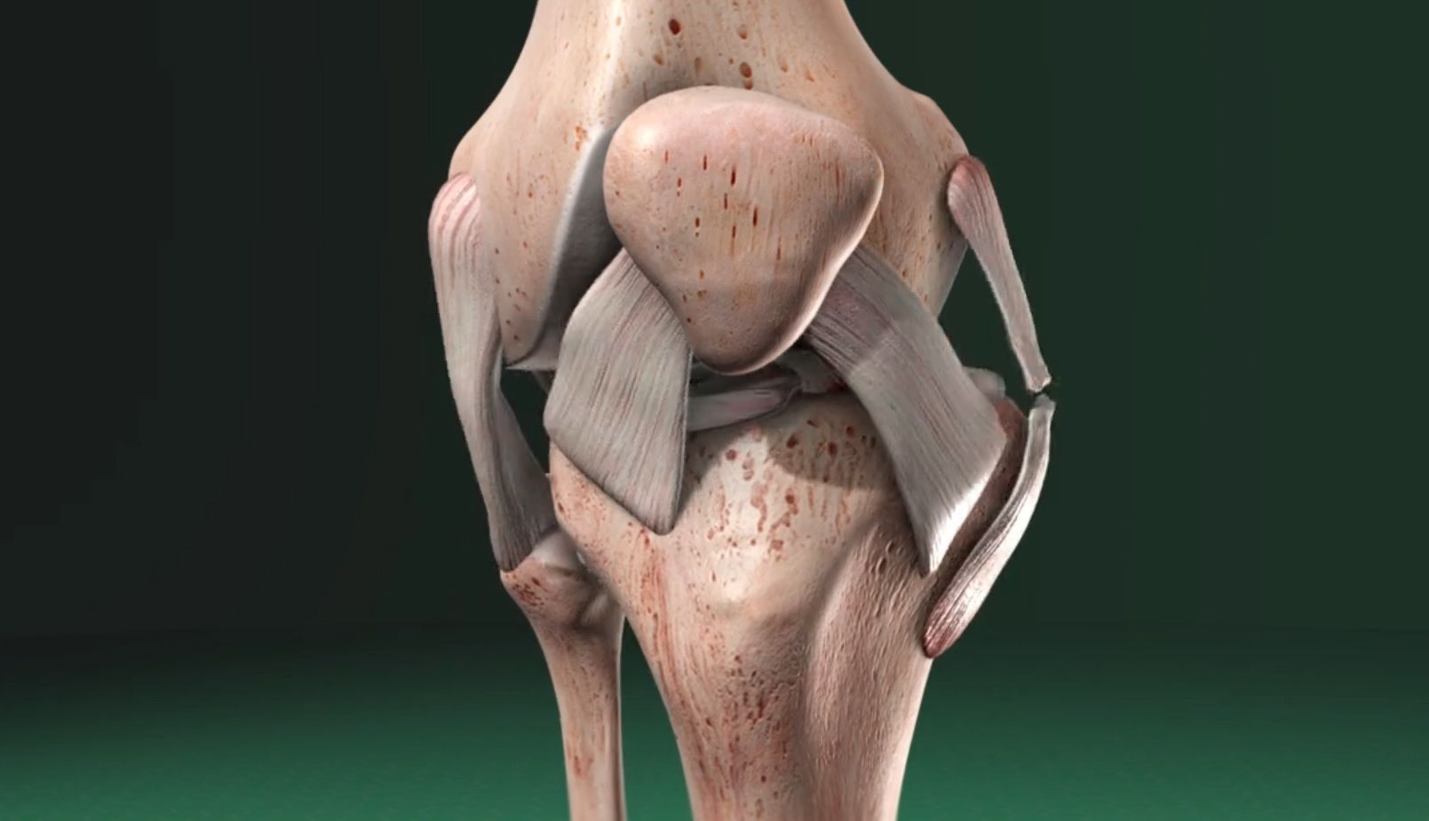
The knee is where three bones meet: the tibia, the femur and the patella. The knee is a “hinge” joint, allowing the leg to bend in only one direction. Here are the main parts of the knee’s anatomy.
Bones
The base of the knee is formed by the tibia, the large bone of the lower leg often called the “shinbone.” The smaller lower leg bone, the fibula, connects to the tibia just below the knee joint. The top of the knee is formed by the femur, the largest bone of the body often called the “thighbone”. The patella, or “kneecap,” covers and protects the front of the knee joint.
Articular Cartilage
The bone surfaces within the knee are covered with a layer of articular cartilage. This tough, smooth tissue protects the bones and allows them to glide smoothly as the knee flexes and extends.
Menisci
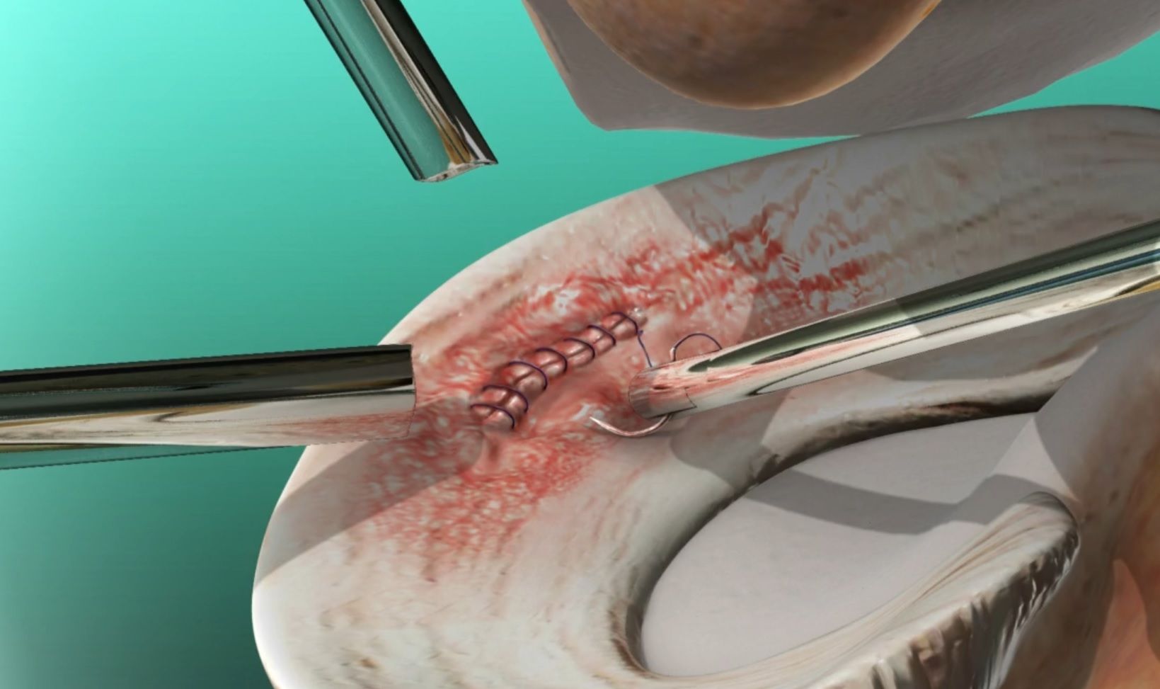
Between the tibia and femur are two thick pads called menisci. Each meniscus is made of cartilage and cushions a condyle, the rounded protrusions on the end of the femur.
Cruciate Ligaments
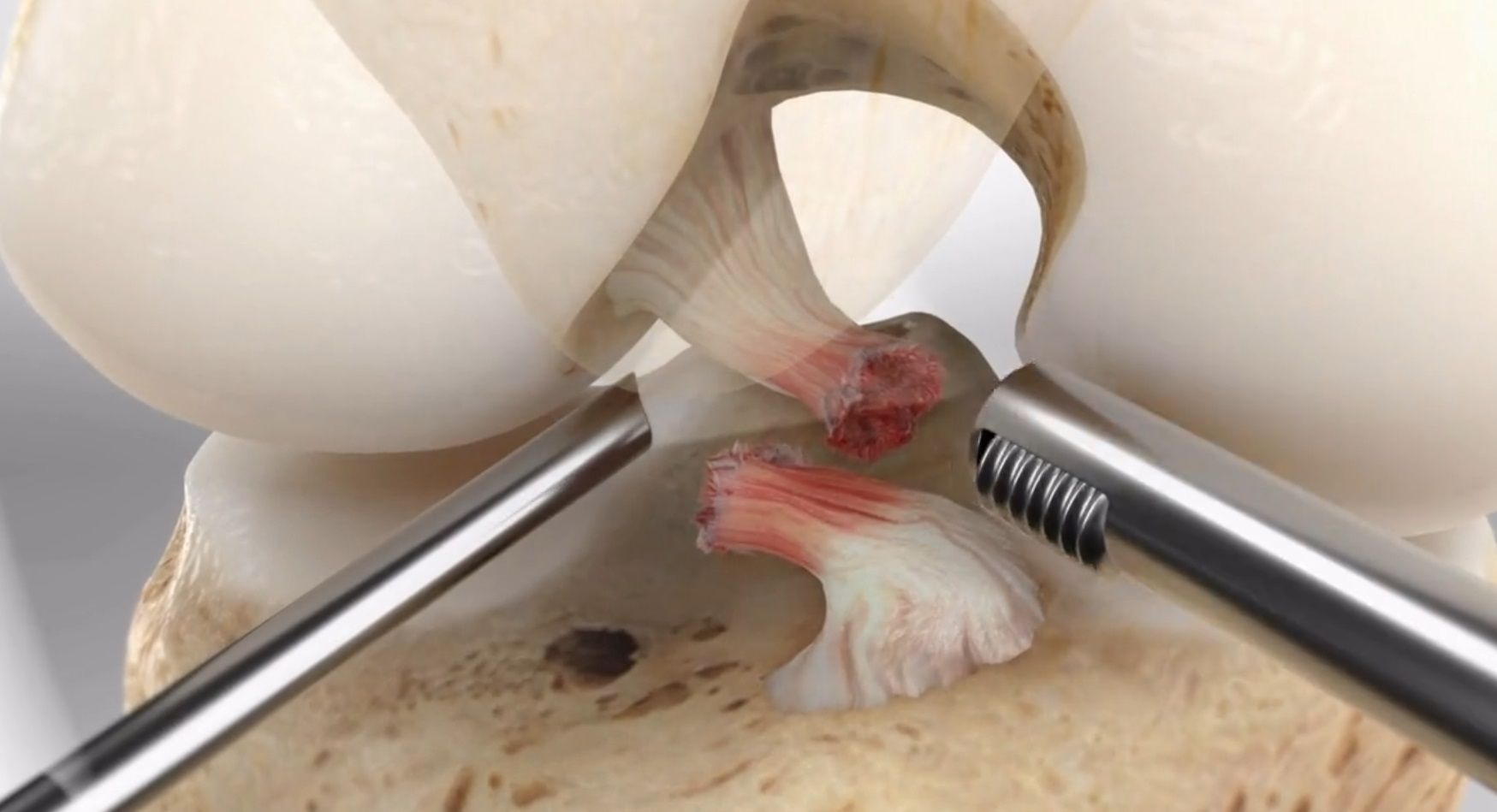
The tibia and the femur are connected together by a pair of strong bands of tissue called cruciate ligaments – the anterior cruciate ligament (ACL) and the posterior cruciate ligament (PCL). These ligaments crisscross each other in the center of the knee, limiting the knee’s rotation and keeping the tibia from slipping forward (ACL) or backward (PCL).
Collateral Ligaments
The collateral ligaments are on the sides of the joint, stabilizing the knee by minimizing side-to-side movement.
Securing the Patella
The patella is secured at the front of the knee by the quadriceps tendon (top) and the patellar ligament (bottom). These connections allow the patella to “float” over the knee as it flexes and extends.
Revised from www.viewmedica.com © Swarm Interactive. Unauthorized duplication is strictly forbidden.
- Category / Anatomy


