Archives: Injury Resources
These are the medical resources that help clients learn more about the various injuries that could be sustained in an accident.
Overview
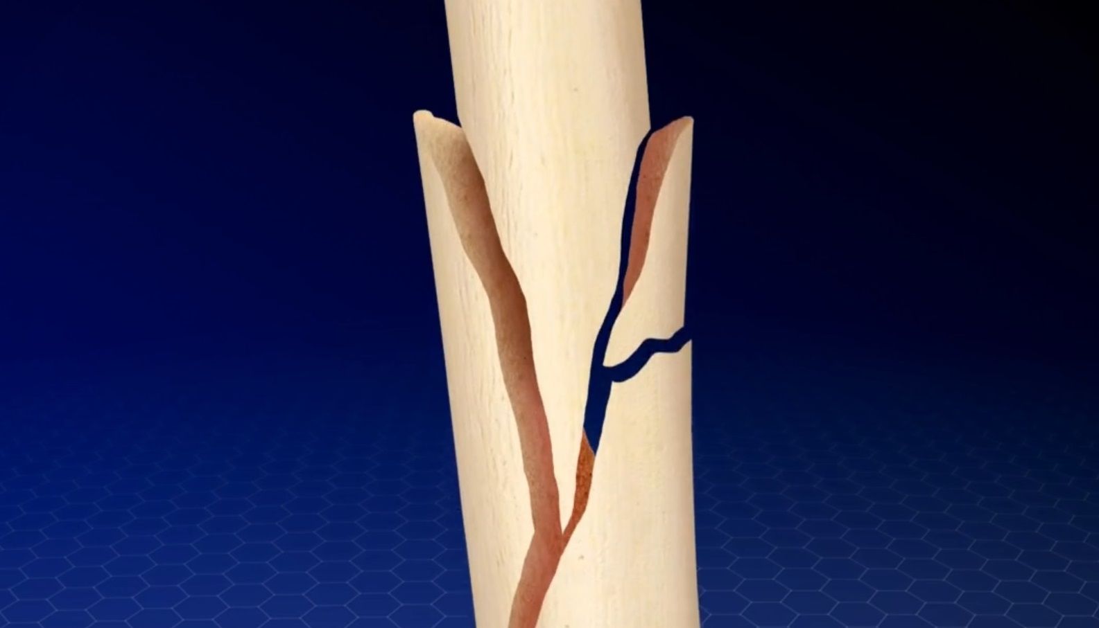
The femur, commonly known as the “thigh bone”, is the largest and strongest bone in your body.
Causes
A direct blow to your thigh can fracture your femur, as can a vehicle crash or fall. Conditions that weaken your bones, like osteoporosis, make a fracture more likely.
Types of Fractures
Femur fractures can happen at the top near the hip socket, along the shaft in the middle of the bone, or down near the knee. Your bone may break in a clean line, at an angle, or in a spiral...
Facet Joint Injections
Overview
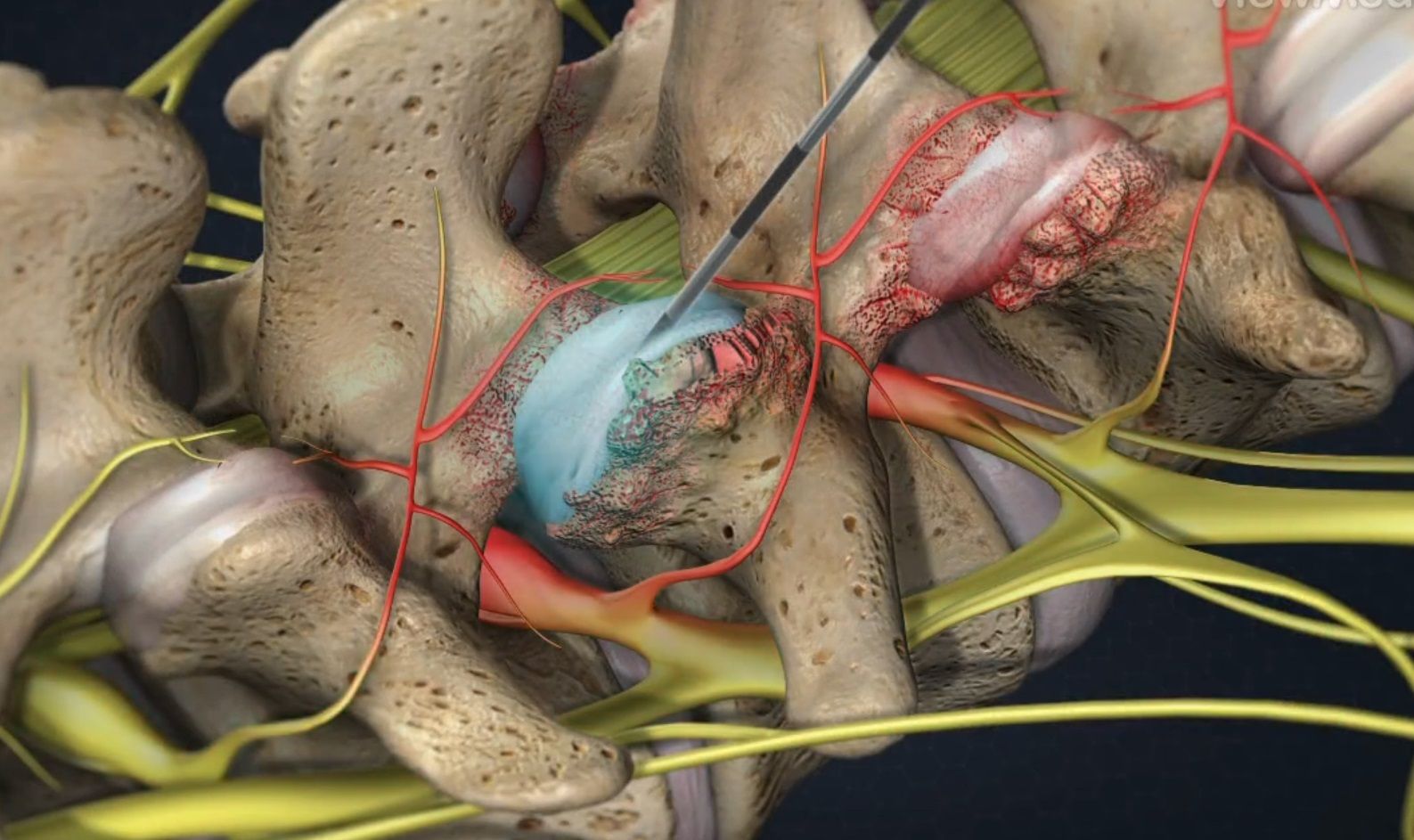
The facet joints are found at the back of spinal vertebrae and can become painfully irritated or inflamed. An injection can relieve inflammation and pain, helping doctors diagnosis the pain’s source.
Skin Numbed
In preparation for the procedure, the physician injects local anesthetic to numb tissue from the skin down to the facet joint.
Placement Confirmed
With the aid of a video x-ray device called a fluoroscope, the physician guides a needle through the numbed tissue and...
Epidural Steroid Injection (ESI) – Lumbar Spine
Overview
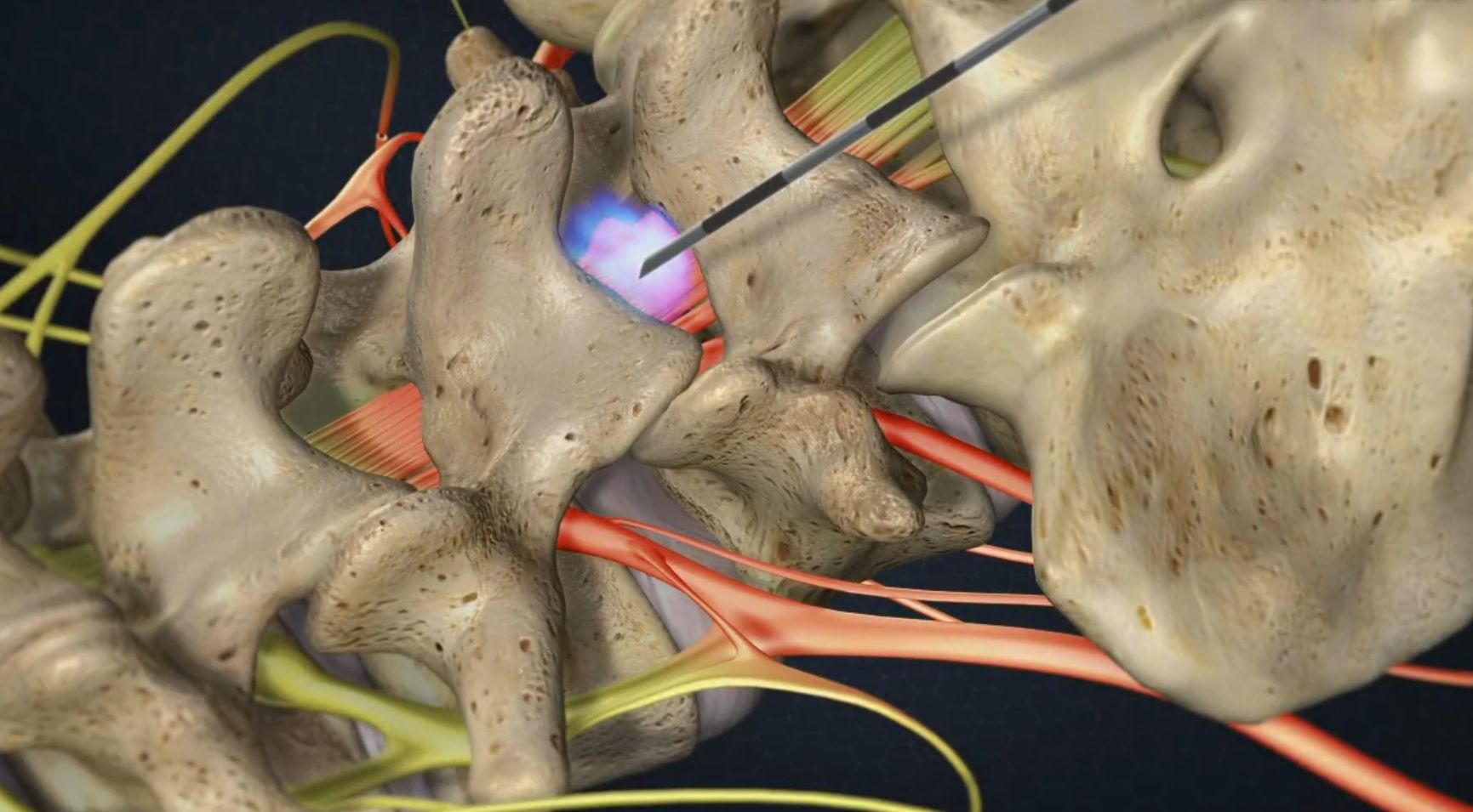
This injection procedure is performed to reduce nerve swelling and inflammation to relieve low back and radiating leg pain.
Patient Positioning
You lie face down with a cushion under the stomach area. This flexes the back to expand the spine and allow the doctor easier access to the epidural space.
Tissue Anesthetized
A local anesthetic is used to numb tissues from the skin all the way down to the surface of the lamina portion of the lumbar vertebra. The physician slides...
Epidural Steroid Injection (ESI) – Cervical Spine
Overview
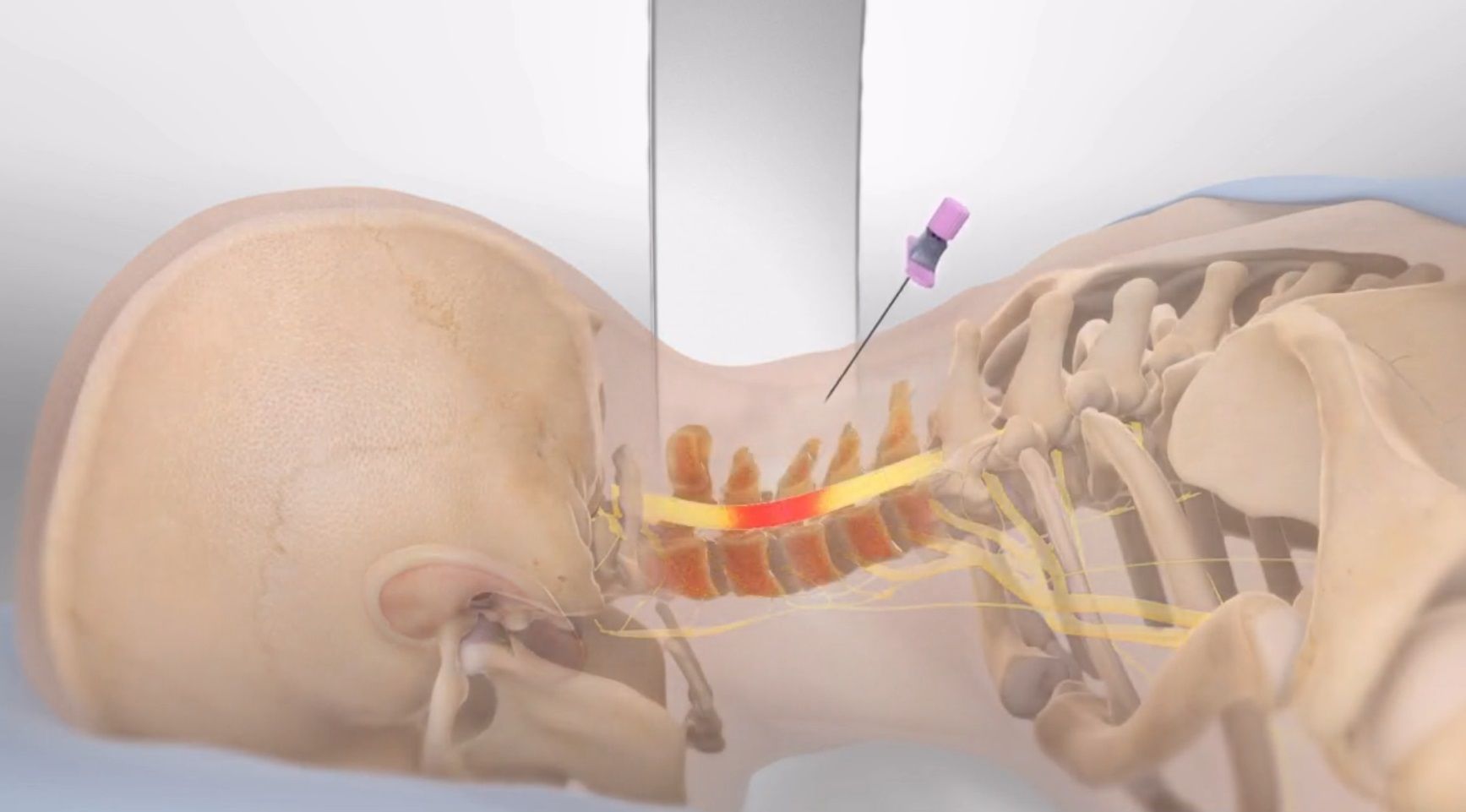
This injection treats painful nerve swelling in your cervical spine (neck), which can be caused by spinal stenosis or a herniated disc pressing on a nerve.
Preparation
To begin, you lie down while local anesthetic is used to numb the injection site.
Placing the needle
A needle is inserted through the numbed tissue. Using a video x-ray device (fluoroscope), the doctor will guide the needle into the epidural space around your inflamed nerve. The doctor may inject contrast...
Electromyography (EMG)
Overview
This is a two-part test of your nerves and muscles. The first part is a nerve conduction study, sometimes called an NCS or NCV (nerve conduction velocity). The NCS measures how well electricity moves through your nerves. The second part is a needle electromyogram (needle EMG), which records the electrical signals your muscles make when you move them. The results can help your doctor find problems linked to certain disorders or conditions.
Nerve Conduction Study
For an NCS/NCV test,...
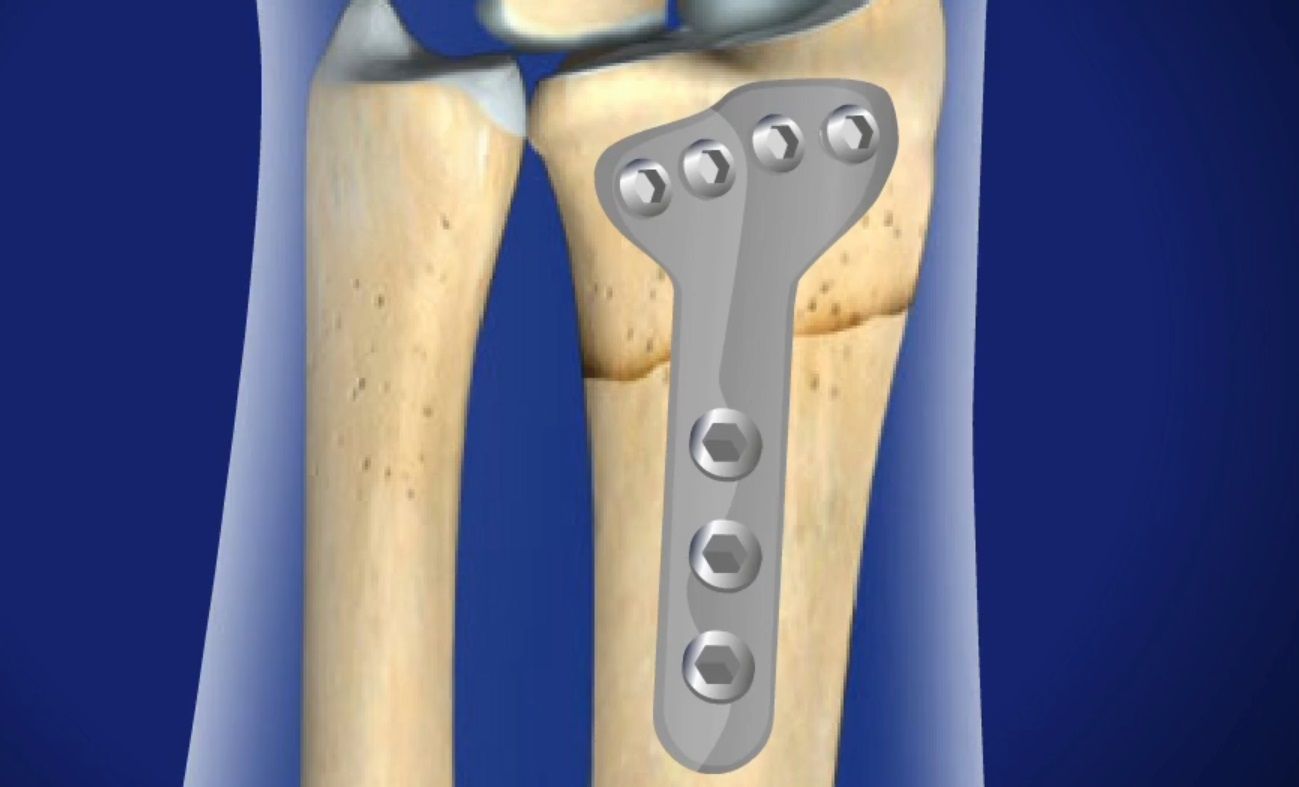
Distal Radius Fracture Repair Surgery
Overview
This open reduction and internal fixation (ORIF) surgery uses screws and a metal implant to stabilize a fracture in the radius near the wrist. The radius is the larger of the two forearm bones.
Preparation
The patient’s arm is positioned with the palm facing up, for optimal surgical access. The area is cleaned, sterilized, and given local anesthesia. Usually the patient is not put to sleep.
Accessing the Fracture
The physician cuts an incision along the forearm to access the region...
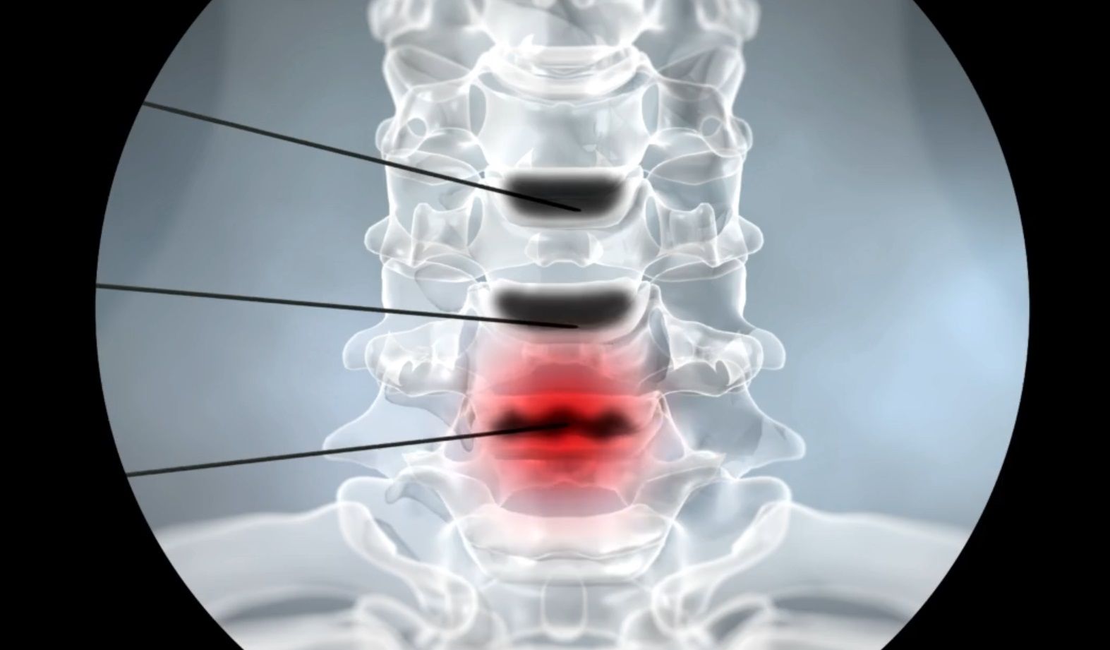
Discography and Discogram
Overview
This procedure, which your doctor may call discography or discogram, helps to identify painful spinal discs. Here’s how it works.
Preparation
You will be relaxed but awake during he procedure. You will not be put to sleep, so that you can give your doctor feedback during the discography. Your neck or back will be numbed with local anesthetic.
Placing the Needles
The doctor will use a video x-ray device called a fluoroscope to guide a needle into each of the suspected painful discs.
Testing...
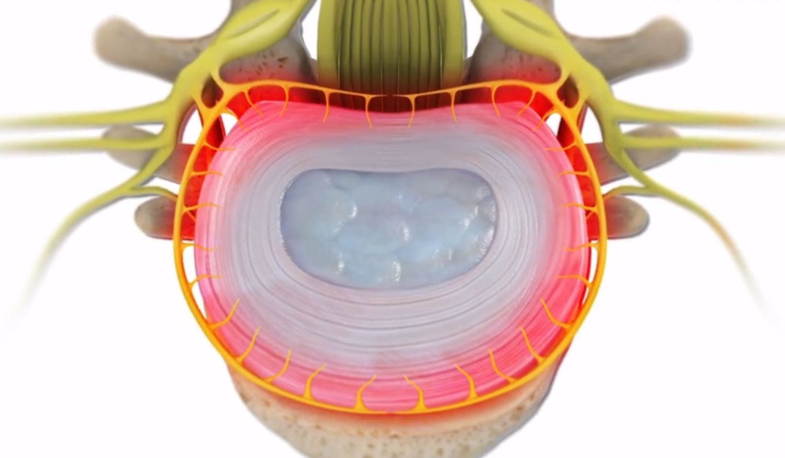
Discogenic Pain
Overview
Spinal discs cushion the spinal bones (vertebrae) above and below them, and allow the spine to move in a variety of directions. These disc have nerves that give the sensation of pain when the disc is injured or weakened. This is called discogenic pain and is different than pain caused by a spinal cord nerve.
Causes and Symptoms
Discogenic pain occurs when the tiny nerves in the outer disc walls are irritated. This happens when the disc is injured and the disc wall tears, allowing fluids...
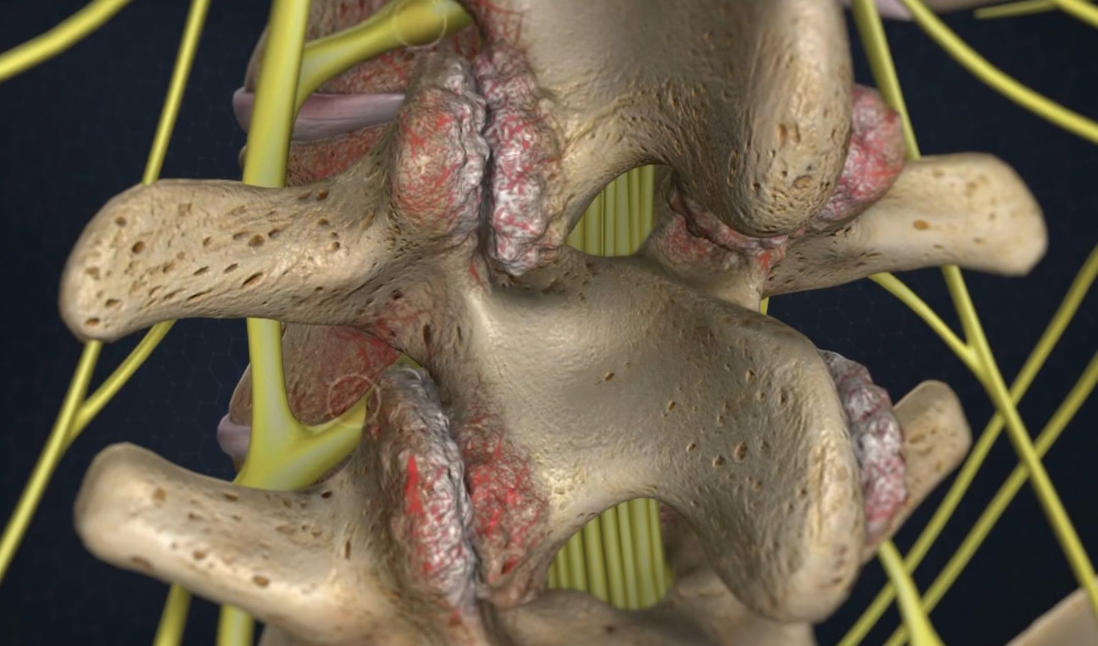
Degenerative Disc Disease
Overview
Vertebral discs act cushion and absorb shock between spinal bones (vertebrae). When discs lose fluid and height, they are said to experience degenerative disc disease. Degeneration happens naturally over time as discs age, but injuries can speed up the degenerative process or make it more severe.
Disc Wall Tears and Scar Tissue
Degenerative disc disease typically starts with small tears in the disc wall (annulus fibrosis). This does not always cause pain. The tears heal with scar...
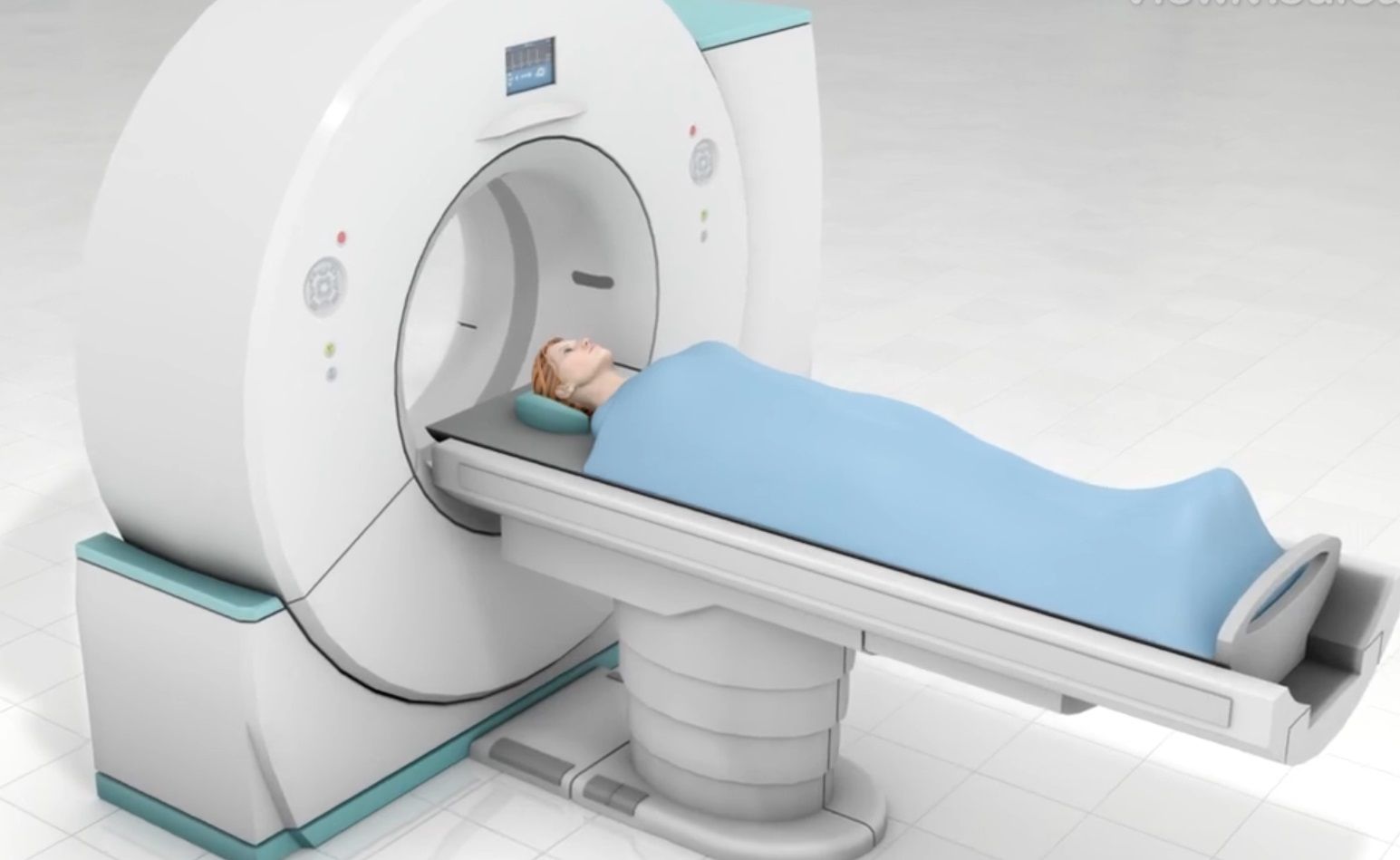
CT Scan (Computed Tomography, CAT Scan)
Overview
This diagnostic scan takes x-ray images from many angles, combining them to show doctors clear cross-section slices of your body parts. A CT scan shows much more than a typical x-ray and depicts structures that MRIs are inadequate to image.
Preparation
You will need to remove your glasses, jewelry, and other metal items before you enter the CT scanner. You will likely be given a hospital gown to wear and may be given medicine to help you relax. Some patients have to drink, or be injected...

